

How to Reverse Low Testosterone: Why Low T Starts in Your Mitochondria
By Courtney Hunt, MD, FACOG | Board-certified OB/GYN

Low testosterone is one of the most searched hormone problems in the world, and one of the most misunderstood. Fatigue, low libido, loss of muscle, weight gain around the middle, brain fog and low motivation send millions of people to a clinic. The usual answer there is to add testosterone back. The number on the lab report goes up. But the reason it fell in the first place is still there.
This guide explains what actually causes low testosterone and why it is best understood as a signal of mitochondrial dysfunction. It covers why replacement therapy does not repair the problem, and what the published research says about reversing low T at its root. Every claim is referenced to peer-reviewed literature so you can read the evidence yourself.
What Causes Low Testosterone? It Starts Before Testosterone Exists
Testosterone is not made from scratch. Every steroid hormone your body produces begins as cholesterol: testosterone, estrogen, progesterone, cortisol, aldosterone and DHEA. The first and slowest step of that cascade is the conversion of cholesterol into pregnenolone.

That conversion is carried out by a single enzyme, P450scc (CYP11A1), and it happens inside your mitochondria (Miller & Auchus, 2011).
Pregnenolone is often called the mother hormone, because every other steroid hormone is built from it. If pregnenolone production slows, everything downstream slows with it. This is why low testosterone so often travels with low DHEA, low progesterone and a flattened cortisol rhythm. These are not several separate problems. They are one upstream problem expressing itself in several places.
Steroid production is the same process repeated in every gland, with local variations: testes, ovaries, adrenals and placenta (Miller & Auchus, 2011). That means the mechanism described here applies to men and to women.
Pregnenolone and the Mitochondria: The Energy Behind Your Hormones
Two things have to happen inside the mitochondria for pregnenolone to be made.
Cholesterol has to be moved. Cholesterol sits on the outer mitochondrial membrane, while the enzyme that converts it sits on the inner membrane. A transport protein called StAR (steroidogenic acute regulatory protein) carries cholesterol across that gap.

This transfer is the rate-limiting, tightly regulated event in hormone production (Stocco, 2001).
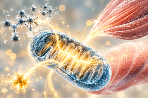
The enzyme needs electrons. CYP11A1 runs on an electron-transfer chain of its own. Adrenodoxin reductase takes electrons from NADPH and passes them to adrenodoxin, an iron-sulfur protein. Adrenodoxin delivers them to CYP11A1, which uses them to cleave cholesterol's side chain (Strushkevich et al., 2011).
Simply stated: making pregnenolone is an energy event. It depends on a functioning, charged, electron-moving inner mitochondrial membrane.
Low Testosterone Is a Sign of Mitochondrial Dysfunction: The Evidence

No mitochondrial energy, no testosterone
Researchers tested this directly in the testosterone-producing Leydig cells of the testes. When they blocked the mitochondrial electron transport chain, cellular energy (ATP) dropped, steroid hormone production was strongly inhibited, and StAR protein levels fell. Mitochondrial membrane potential, mitochondrial ATP synthesis and mitochondrial pH were each required.
Their conclusion was that mitochondria must be energized, polarized and actively respiring to make steroid hormones (Allen et al., 2006; Hales et al., 2005).
Inflammation and oxidative stress shut the factory down
The same research group showed that oxidative stress collapses the mitochondrial membrane potential. It blocks StAR at every stage: its production, its import into the mitochondria, and its ability to move cholesterol (Diemer et al., 2003). In living animals, bacterial endotoxin, a potent inflammatory trigger, produced reactive oxygen species that damaged Leydig cell membranes. Mitochondrial membrane potential collapsed and testosterone production shut down (Allen et al., 2004).
Importantly, when the oxidative stress was removed, StAR and hormone production recovered (Diemer et al., 2003). The cells were not destroyed. They were switched off. That is the foundation of reversal.
In humans, low T tracks with low mitochondrial function
A Massachusetts General Hospital study of 60 men found that testosterone levels correlated with insulin sensitivity and with aerobic capacity (VO2max). The strongest correlation was with expression of the oxidative phosphorylation genes in skeletal muscle, the genes that run mitochondrial energy production. this is why I teach correcting insulin resistance and improving VO2 max in my classes.
Men with low testosterone had three times the prevalence of metabolic syndrome (Pitteloud et al., 2005a). A companion study found that as insulin resistance rises, testosterone secretion from the Leydig cells themselves falls (Pitteloud et al., 2005b). The defect is in the hormone-producing cell, not only in the brain's signal.
Aging works through the same pathway
Age-related low testosterone results from reduced production by Leydig cells, not from losing them (Beattie et al., 2015). Aged Leydig cells carry significantly more superoxide, a mitochondrial free radical. The activity of StAR, CYP11A1 and the downstream hormone-building enzymes all declines (Beattie et al., 2015; Zirkin & Tenover, 2012). The "normal aging" of testosterone is, at the cellular level, accumulated oxidative damage to the mitochondrial hormone machinery.
Why Testosterone Replacement Doesn't Fix Low T
Testosterone replacement therapy (TRT) raises blood testosterone. It does not restore StAR transport, adrenodoxin electron flow, CYP11A1 activity, mitochondrial membrane potential or insulin sensitivity. The upstream energy problem that lowered testosterone is left exactly where it was.
Replacement also changes the system it is added to. When testosterone is given from outside, the pituitary lowers its signal (LH) to the testes. The testes then make less of their own testosterone, intratesticular testosterone falls, and sperm production is impaired (Beattie et al., 2015). The Endocrine Society's clinical guideline makes three relevant recommendations (Bhasin et al., 2018):
• Evaluate further to determine the cause of low testosterone before treating.
• Do not use testosterone therapy in men planning fertility in the near future.
• Do not use testosterone as a way to improve blood sugar control.
One finding is worth knowing. In aging rats, suppressing the testes with testosterone implants for a long period protected the Leydig cells from age-related decline (Chen & Zirkin, 1999). The explanation is that the work of making hormones itself generates oxidative stress over time. That supports the mitochondrial model rather than contradicting it: replacement rests the factory. It does not repair it.
Testosterone therapy has real, appropriate uses, including true structural disease of the testes or pituitary. But for most people with low T, the more durable question is not "How do I add testosterone?" It is "Why did my mitochondria stop making it?"
Functional Low Testosterone Is Reversible
Most middle-aged and older men with low testosterone have no structural disease of the brain, pituitary or testes. Their low testosterone is functional, and potentially reversible. Leading endocrinologists recommend treating the underlying causes first, or alongside therapy (Grossmann & Matsumoto, 2017). A 2023 clinical review in the Journal of Clinical Endocrinology & Metabolism goes further. It advises considering a trial of testosterone only after at least 6 to 12 months of addressing reversible causes, including vigorous lifestyle change (Grossmann et al., 2023).
How to Reverse Low Testosterone at the Root

The research points to the same targets: restore mitochondrial energy, reduce inflammation and oxidative stress, and correct the metabolic signals that suppress hormone production.
1. Reverse insulin resistance
Insulin resistance is directly associated with reduced testosterone secretion from the testes (Pitteloud et al., 2005b), and both track with mitochondrial function (Pitteloud et al., 2005a). Restoring insulin sensitivity is the central lever. Build meals around animal protein: beef, lamb, eggs, fish and organ meats. These supply complete protein along with cholesterol, the raw material for every steroid hormone.
2. Lose visceral fat
A meta-analysis of weight-loss studies found that losing weight raises both total and free testosterone. The amount of weight lost was the strongest predictor of how much testosterone rose (Corona et al., 2013). Fat tissue is not passive: it drives inflammation and converts testosterone to estrogen.
3. Lower inflammation and oxidative stress
Inflammation and reactive oxygen species collapse the mitochondrial membrane potential and switch off StAR (Diemer et al., 2003; Allen et al., 2004). Identifying and removing chronic inflammatory triggers takes the brake off. These include gut-derived endotoxin, chronic infection and toxic exposures.
4. Protect sleep
In healthy young men, one week of sleep restricted to five hours a night lowered daytime testosterone by 10–15% (Leproult & Van Cauter, 2011). Testosterone production is concentrated during sleep, and the mitochondria repair during sleep.
5. Review medications
Some medications suppress the hormone axis. Where appropriate, stopping the offending medication is part of the guideline-supported approach to functional low T (Grossmann & Matsumoto, 2017). Do this only with your prescribing physician.
6. Test correctly, and retest
Diagnosis requires symptoms plus consistently low morning testosterone on repeat testing (Bhasin et al., 2018). To confirm that reversal is happening at the source, track these over time:
• Total and free testosterone
• SHBG
• LH
• Fasting insulin
• Markers of inflammation
Low Testosterone in Women
Women make testosterone in the ovaries and adrenals through the same cholesterol-to-pregnenolone pathway, beginning in the same mitochondria (Miller & Auchus, 2011). In women, low testosterone often accompanies low progesterone, fatigue, low libido and loss of muscle tone, particularly in perimenopause. The same upstream logic applies. Restore the mitochondrial energy that makes the mother hormone, and the downstream hormones have the raw material they need.
Frequently Asked Questions
Can low testosterone be reversed naturally?
In most cases of functional low testosterone, yes. Research shows functional low T is potentially reversible. Treating underlying causes such as obesity, insulin resistance and comorbidities raises testosterone (Grossmann & Matsumoto, 2017; Corona et al., 2013).
What is the root cause of low testosterone?
At the cellular level, testosterone production depends on mitochondria converting cholesterol to pregnenolone. Insulin resistance, inflammation, oxidative stress and aging all impair this mitochondrial step (Allen et al., 2006; Pitteloud et al., 2005a; Beattie et al., 2015). Again thank youhis is why I take a deep dive into teaching the correction of these things.
Does testosterone replacement therapy fix low testosterone?
TRT raises blood levels but does not repair the mitochondrial cause. It also suppresses LH, lowering your own testosterone production and sperm production (Beattie et al., 2015; Bhasin et al., 2018).
What is pregnenolone and why does it matter for testosterone?
Pregnenolone is the first hormone made from cholesterol, inside the mitochondria. It is the precursor to testosterone, estrogen, progesterone, cortisol and DHEA, and its production is the rate-limiting step for all steroid hormones (Miller & Auchus, 2011).
Does losing weight increase testosterone?
Yes. A meta-analysis found that weight loss raises both total and free testosterone, in proportion to the amount of weight lost (Corona et al., 2013).
Is low testosterone linked to insulin resistance?
Yes. Testosterone correlates with insulin sensitivity and with mitochondrial gene expression. Rising insulin resistance is associated with lower testosterone secretion by the testes (Pitteloud et al., 2005a, 2005b).
How long does it take to raise testosterone naturally?
Clinical guidance suggests addressing reversible causes for at least 6 to 12 months before considering a trial of testosterone therapy (Grossmann et al., 2023). In my classes I see things reverse clinically in the first three months. While this is anecdotal it is consistent with the literature.
The Bottom Line
Low testosterone is less a testosterone deficiency than an energy deficiency. It is a downstream readout of what is happening in your mitochondria. Adding testosterone silences the signal. Restoring mitochondrial function addresses the cause: reverse insulin resistance, lower inflammation, protect sleep and supply the right raw materials.
Energy over inflammation is the basic equation.
This article is for education and is not a substitute for individualized medical care. Do not stop or change prescribed hormone therapy without your physician.
© Courtney Hunt, MD, 2026
References
1 Miller WL, Auchus RJ. The molecular biology, biochemistry, and physiology of human steroidogenesis and its disorders. Endocr Rev. 2011;32(1):81–151.
2 Stocco DM. StAR protein and the regulation of steroid hormone biosynthesis. Annu Rev Physiol. 2001;63:193–213.
3 Strushkevich N, et al. Structural basis for pregnenolone biosynthesis by the mitochondrial monooxygenase system. PNAS. 2011;108(25):10139–10143.
4 Allen JA, et al. Energized, polarized, and actively respiring mitochondria are required for acute Leydig cell steroidogenesis. Endocrinology. 2006;147(8):3924–3935.
5 Hales DB, et al. Mitochondrial function in Leydig cell steroidogenesis. Ann N Y Acad Sci. 2005;1061:120–134.
6 Diemer T, et al. Reactive oxygen disrupts mitochondria in MA-10 tumor Leydig cells and inhibits StAR protein and steroidogenesis. Endocrinology. 2003;144(7):2882–2891.
7 Allen JA, et al. Bacterial endotoxin lipopolysaccharide and reactive oxygen species inhibit Leydig cell steroidogenesis via perturbation of mitochondria. Endocrine. 2004;25(3):265–275.
8 Pitteloud N, et al. Relationship between testosterone levels, insulin sensitivity, and mitochondrial function in men. Diabetes Care. 2005;28(7):1636–1642. (2005a)
9 Pitteloud N, et al. Increasing insulin resistance is associated with a decrease in Leydig cell testosterone secretion in men. J Clin Endocrinol Metab. 2005;90(5):2636–2641. (2005b)
10 Beattie MC, et al. Leydig cell aging and hypogonadism. Exp Gerontol. 2015;68:87–91.
11 Zirkin BR, Tenover JL. Aging and declining testosterone: past, present, and hopes for the future. J Androl. 2012;33(6):1111–1118.
12 Chen H, Zirkin BR. Long-term suppression of Leydig cell steroidogenesis prevents Leydig cell aging. PNAS. 1999;96(26):14877–14881.
13 Bhasin S, et al. Testosterone therapy in men with hypogonadism: an Endocrine Society clinical practice guideline. J Clin Endocrinol Metab. 2018;103(5):1715–1744.
14 Grossmann M, Matsumoto AM. A perspective on middle-aged and older men with functional hypogonadism: focus on holistic management. J Clin Endocrinol Metab. 2017;102(3):1067–1075.
15 Grossmann M, Jayasena CN, Anawalt BD. Approach to the patient: men ≥50 years with low serum testosterone. J Clin Endocrinol Metab. 2023;108(9):e871–e884.
16 Corona G, et al. Body weight loss reverts obesity-associated hypogonadotropic hypogonadism: a systematic review and meta-analysis. Eur J Endocrinol. 2013;168(6):829–843.
17 Leproult R, Van Cauter E. Effect of 1 week of sleep restriction on testosterone levels in young healthy men. JAMA. 2011;305(21):2173–2174.


Comments